
Automation has transformed urinalysis by improving throughput, standardizing particle counts, and reducing reliance on labor-intensive manual microscopy. But even high-performing automated systems have blind spots. This means that the practical question is not whether manual urine sediment examination still matters, but when does it add meaningful clinical value?
Current guidance favors a selective approach. Laboratories should establish predefined criteria for microscopic review after automated particle analysis. Review criteria should reflect the analytical performance of the specific analyzer, the patient population, and clinical needs (1, 2). Because analyzer performance differs, review rules should be validated locally rather than adopted from another laboratory.
Know the analyzer’s blind spots
Automated systems are useful for standardized enumeration of common particles such as red blood cells (RBCs) and white blood cells (WBCs) (1, 3), but limitations become more important when detailed morphology is required. Dysmorphic RBCs, acanthocytes, pathologic casts, renal tubular epithelial cells, atypical cells, unusual crystals, and other difficult-to-classify particles may require visual confirmation (1, 2).
A 2025 comparison with reference microscopy found that automated systems had sensitivities below 50% for pathologic casts, lipiduria, and renal tubular epithelial cells and 62.3% for dysmorphic RBCs (4). In the multicenter ADORE study, the two higher-performing systems for pathologic-cast detection had sensitivities of 58.6% and 69.6%, and 90% of samples required manual on-screen review to achieve acceptable performance for dysmorphic RBC and cast detection (5). Selected published performance data are summarized in Figure 1.
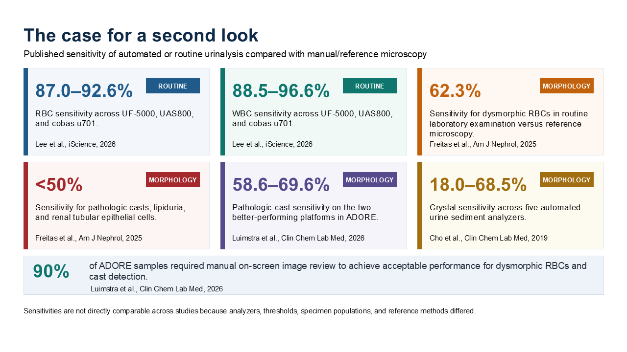
Performance also varies by technology and particle type. The UF-5000 flow cytometry analyzer performed better than digital-imaging systems for bacteriuria and mucus threads but less well for crystals (3). Cast and crystal detection also varied across platforms and cutoff settings (6). Unusual organisms and morphologies can challenge automated classification; published yeast-like and mold-like cases illustrate why findings that do not match expected morphology deserve closer review (7, 8).
What should trigger review?
A practical algorithm should focus on findings for which review could change reporting or interpretation. Common triggers include instrument flags or uncertain classifications, chemistry-particle discordance, and suspicion of clinically important morphology. Examples include pathologic or unclassified casts, dysmorphic RBCs, nonsquamous epithelial cells, yeast, unusual crystals, unidentified particles, substantial dipstick blood with few RBCs, positive leukocyte esterase with few WBCs, or marked turbidity without corresponding particles.
Dipstick thresholds can help prioritize review but should not serve as the sole safeguard. In 503 predominantly nephrology specimens, protein ≥1+ or blood ≥2+ identified nephropathy-associated sediment findings with 71.2% sensitivity and 73.9% specificity (9). These thresholds can enrich specimens for review but would miss important findings if used alone.
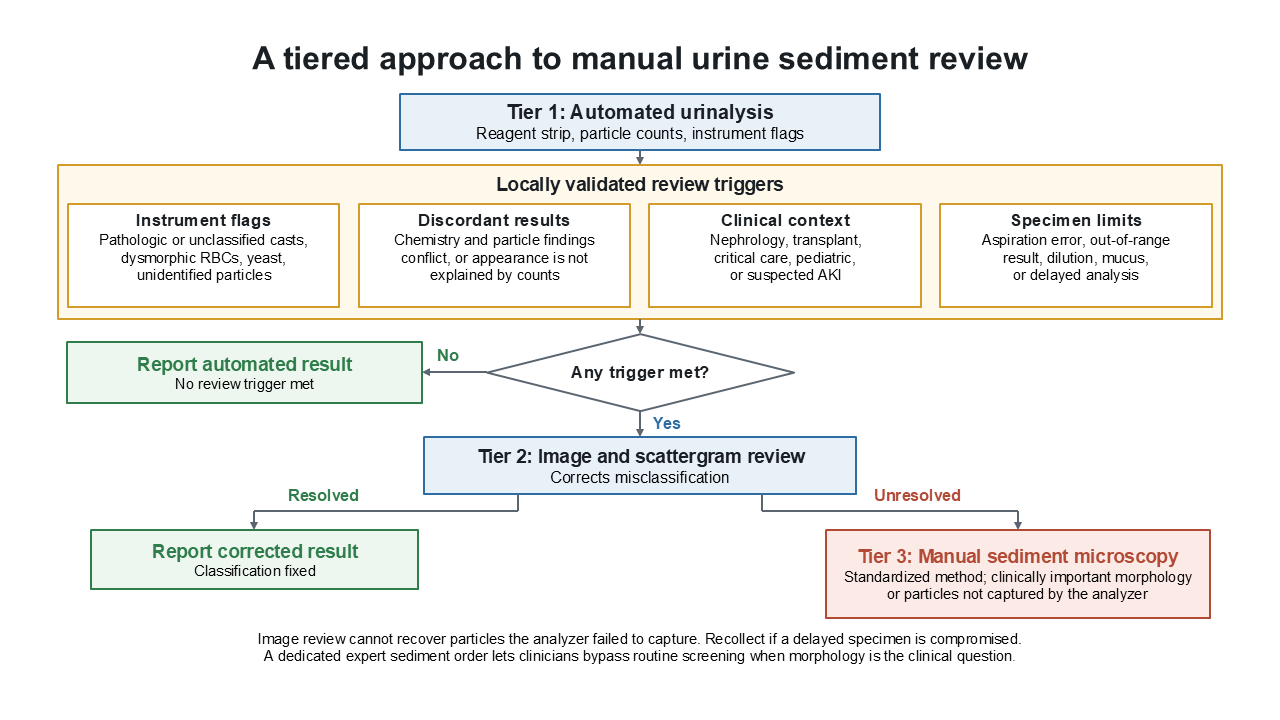
When reviewing automated findings, it’s important to keep in mind that image review and manual sediment microscopy are not interchangeable. Image review may correct classification errors but cannot recover particles that were not adequately captured by the analyzer. If image review does not resolve a misclassification, manual sediment microscopy should be turned to next.
Additionally, discordance does not necessarily indicate analyzer failure. A heme-positive dipstick with few or no intact RBCs may reflect hemoglobin, myoglobin, or RBC lysis. Automated systems may also underestimate hematuria in glomerular disease (10). The goal is to resolve the discrepancy before reporting an internally inconsistent urinalysis.
Clinical setting and specimen quality
Review thresholds may reasonably be lower when clinically important renal disease is suspected, including acute kidney injury or renal transplant dysfunction. It may also be appropriate to bypass automated screening and go straight to expert sediment microscopy when detailed morphology is the clinical question (1, 2, 11).
Dysmorphic RBCs should be interpreted with proteinuria, renal function, pathologic casts, and clinical findings rather than in isolation (12). In 131 patients undergoing kidney biopsy, renal tubular epithelial cells and urinary lipids were associated with clinically relevant histologic lesions (13).
Specimen quality also matters. Delayed analysis and specimens with abundant mucus, crystals, debris, or other artifacts can compromise sediment evaluation. Cells and casts may deteriorate over time; when specimen integrity is poor, recollection may be preferable to interpreting a compromised sample (1, 11, 12). Clinical and Laboratory Standards Institute guideline PRE05 provides guidance on urine collection, handling, transport, and specimen quality (14).
Use a tiered approach
Not every abnormal automated result needs manual microscopy. A tiered process preserves efficiency while directing expertise where it matters most (Figure 2). Automated analysis should handle particles it measures reliably. Instrument images and scattergrams can resolve straightforward classification questions. Manual sediment microscopy should be reserved for unresolved findings, clinically important morphology, or discordance that cannot otherwise be explained.
Manual microscopy must also be standardized. Specimen mixing, centrifugation, sediment concentration and resuspension, examined volume, microscopy technique, terminology, and reporting units can all introduce variability (1, 2, 11). Competency assessment should include low-frequency, high-consequence findings rather than only common RBCs and WBCs.
The best review rule is not “microscopy for every abnormal urinalysis.” The best rule is a locally validated safety net built around the analyzer’s known limitations, chemistry-particle concordance, specimen quality, and clinical context. The goal is not to examine more specimens manually, but to identify those in which expert microscopy can make a difference.
Jingcai Wang, MD, PhD, DABCC, NRCC, ASCP, FADLM, is a clinical assistant professor at The Ohio State University in Columbus, assistant director of clinical chemistry and immunology at Nationwide Children’s Hospital, and a College of American Pathologists/CLIA laboratory director for multiple neonatal intensive care unit laboratory sites. +Email: [email protected]
Read the full November-December issue of CLN.
References
- Pons-Belda OD, Ferri MJ, Almería-Lafuente A, et al. Recommendations for the standardization and clinical reporting of urinary sediment in the clinical laboratory. Adv Lab Med 2026; doi:10.1515/almed-2026-0070.
- Kouri TT, Hofmann W, Falbo R, et al. The EFLM European urinalysis guideline 2023. Clin Chem Lab Med 2024;62:1653-786.
- Lee J, Choi H, Jang H, et al. Comparative evaluation of three automated urine sediment analyzers excluding cutoff-related trade-offs. iScience 2026;29:114438.
- Freitas PAC, da Silva YDS, Poloni JAT, et al. The clinical impact of urinalysis screened by automated microscopy compared to reference manual analysis. Am J Nephrol 2025;56:178-86.
- Luimstra JJ, Manz XD, Koçer RG, et al. ADORE: The performance characteristics of five automated platforms in the analysis of urinary dysmorphic erythrocytes and pathological casts. Clin Chem Lab Med 2026;64:421-31.
- Cho J, Oh KJ, Jeon BC, et al. Comparison of five automated urine sediment analyzers with manual microscopy for accurate identification of urine sediment. Clin Chem Lab Med 2019;57:1744-753.
- Wang J, Nicol KK, Pyle-Eilola AL. Budding yeast that won’t stain. Clin Chem 2021;67:1432-3.
- Kara S, Oyeniran SJ, Kahwash SB, et al. A mold-like organism in urine sediment. Clin Chem 2026;72:321-2.
- Freitas PAC, da Silva YDS, Poloni JAT, et al. Dipstick proteinuria and hematuria as triggers for manual microscopic review in nephrology patients. J Clin Med 2025;14:4522.
- Yang WS. Automated urine sediment analyzers underestimate the severity of hematuria in glomerular diseases. Sci Rep 2021;11:20981.
- Cavanaugh C, Perazella MA. Urine sediment examination in the diagnosis and management of kidney disease: Core curriculum 2019. Am J Kidney Dis 2019;73:258-72.
- Pons-Belda OD, Ferri MJ, Almería-Lafuente A, et al. Recommendations for optimizing the use of dysmorphic red blood cell counts in the clinical laboratory. Adv Lab Med 2026;7:171-8.
- Navarro D, Fonseca NM, Ferreira AC, et al. Urinary sediment microscopy and correlations with kidney biopsy: Red flags not to be missed. Kidney360 2023;4:32-40.
- Clinical and Laboratory Standards Institute. Processes for the collection of urine specimens. 1st ed. CLSI guideline PRE05. 2024.