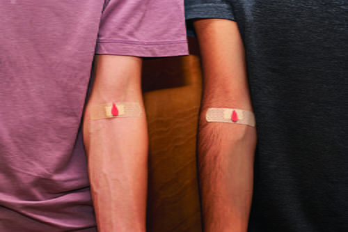
Most laboratory errors occur in the preanalytical phase. Studies have demonstrated that 60−70% of errors occur prior to specimens being received in the laboratory (1). Most of these errors are often be attributed to the practice of phlebotomy. Phlebotomy, defined simply as the act of withdrawing blood from a patient using a needle, is a critical first step in the process of laboratory testing.
Despite its importance for quality laboratory testing, the significance of correct phlebotomy is often overlooked. A survey by the European Federation of Clinical Chemistry and Laboratory Medicine (EFLM) found that phlebotomy training was required curriculum for only 21% of nursing programs and 32% of technologist programs (1). In the U.S., requirements for performing phlebotomy vary widely. For example, some states, such as California, require licensing to perform phlebotomy. In contrast, states such as Missouri require only on-the-job training and no formal certification.
In many U.S. hospitals, nursing staff is permitted to draw blood with minimal training and no formal certification in phlebotomy. Although there is little recent data, a CAP Q Probes survey from 1991 found that only 17% of the 393 intuitions surveyed required a phlebotomy training course (2).
Although considering the impact of improper phlebotomy, many laboratorians jump to hemolysis as the primary negative outcome. Indeed, hemolysis is an important clinical problem and one of the most frequent preanalytical errors. However, a narrow focus on hemolysis overlooks the potential for errors at any step in the process, and the potential affect on the patient.
The entire process of phlebotomy and best practice are discussed in detail in the Clinical & Laboratory Standards Institute (CLSI) document GP47 (3). Each step has important processes and safeguards built in place that protect the patient and the phlebotomist while procuring an acceptable specimen for clinical testing.
PATIENT AND SPECIMEN IDENTIFICATION
Correct patient identification is crucial. This is typically completed by asking the patient their full name and date of birth and then confirming these details with their arm band (when hospitalized) or photo ID (if not hospitalized). These sources are then compared to the test request and/or sample labels. Errors in this phase of phlebotomy lead to wrong blood in tube errors; patient specimens are drawn into tubes meant for other patients and the results from testing attributed to the wrong patient.
The frequency of wrong blood in tube errors has decreased considerably with the implementation of positive patient identification (PPID), which is used to label the specimen containers at the bedside. However, this technology is infrequently used in outpatient settings and has not been universally adopted. Labs have also attempted to mitigate wrong-blood-in-tube errors by implementing delta checks. A delta check uses the lab information system to compare results from a previous sample to the current sample for an individual patient. A set of rules are applied with the goal of finding changes in an analyte that is unlikely to occur physiologically. While once state of the art, delta checks are often thought of as unnecessary in the PPID era.
PHLEBOTOMY TECHNIQUE
Once the correct patient has been identified, the phlebotomist must prioritize site selection. Most often, median cubital veins in the forearm are used with the lateral, outer veins considered peripherally to avoid nerves that are closer to these veins. The appropriate needle must be selected, generally a 21G or 23G needle, with smaller needles (higher gauge) increasing the likelihood of hemolysis. The site must be appropriately cleaned with 70% alcohol, according to the World Health Organization (WHO), and allowed to dry to reduce likelihood of infection or contamination of blood culture bottles with skin flora. A tourniquet must then be applied only for one minute to avoid pooling of analytes and patient discomfort/ harm, and the patient told not to pump their fist, which can increase the release of potassium locally, causing an artificially high result.
For hospitalized patients, it is common for specimens to be collected from indwelling venous or arterial catheters. While convenient for the hospital staff and often preferred by patients (as opposed to fresh venipuncture), this practice is associated with increased blood culture contamination rates and hemolysis.
Hospitals and laboratories should consider the potential benefits to patients with the potential hazards when drawing from indwelling lines. If a previous indwelling line for vascular access is used, all intravenous (IV) fluids must be stopped, the line flushed with 0.9% sodium chloride, and a proportion (typically 1 mL) of blood should be discarded (usually in a red top serum tube) prior to collecting blood into specimen containers designated for laboratory testing.
IV fluid contamination is a relatively common problem when blood is collected from an indwelling line that is not appropriately flushed. Delta checks are commonly used by laboratories to assess for IV fluid contamination but are nonspecific and lack sensitivity for this purpose. Recently, there has been a rise in publications assessing novel tools for distinguishing IV fluid contamination.
A recent manuscript from Yale demonstrated multianalyte delta checks were able to increase the sensitivity for IV fluid contamination (4). Briefly, the authors used wet-bench experiments to define rules in which 10% contamination with IV fluid would be detected. For example, a rule implemented in which chloride increased by 7.7 mmol/L, potassium decreased by 0.7 mmol/L, and calcium decreased by 1.7 mg/dL was able to accurately detect patients with presumed normal saline contamination.
Another recent manuscript in Clinical Chemistry used unsupervised machine learning and a dimension reduction technique to increase the sensitivity for detection of IV fluid contamination (5). Using this approach, the authors demonstrated an estimated positive predictive value of 78% relative to manual chart review by trained laboratory directors and the model was able to accurately detect almost three times more contaminated specimens than technologists during their routine workflows. Optimistically, these studies will soon lead to novel tools for laboratories to detect contamination in real-time.
TUBE TYPES
A common error labs encounter is specimens collected into tubes with the wrong anticoagulant, or tubes that have been drawn in the wrong order. It is crucial that specimens are not contaminated with anticoagulants that can impact testing. The typical order of draw is: 1. blood culture bottles, 2. serum tubes (red top), 3. sodium citrate tubes (blue top, coagulation testing is very sensitive to other anticoagulants), 4. lithium heparin tubes (green top, often switched with K2 EDTA, resulting in falsely increased potassium and decreased calcium), 5. K2 EDTA tubes (purple or pink top), and 6. others, including gray top tubes for glucose and lactate testing.
When tubes are drawn out of order, a small amount of anticoagulant can be transferred to the subsequent tube, potentially affecting testing. For example, it is not uncommon for hospitalized patients to have prolonged protime (PT) and partial thromboplastin time (PTT) due to anticoagulant therapy. If a blue top citrated tube is drawn immediately after a green top tube, both PT and PTT likely will be artificially prolonged by lithium heparin contamination. Deciphering these contaminants from a patient on anticoagulant therapy is incredibly difficult for a laboratory technologist and potentially for a physician.
SPECIMEN TRANSPORT
A final consideration for specimen collection is transportation of the specimen to the laboratory. Within hospitals, pneumatic tube systems are commonly used to transport specimens. While effective and rapid, they also exert considerable forces on the specimen resulting in hemolysis and potentially other pre-analytical errors.
Laboratories should consider validating the pneumatic tube system and the potential impact on patient specimens, as manufacturers do not commonly do this. While external laboratory transport is likely less susceptible to traumatic hemolysis, temperature and time from collection may impact testing and is not commonly considered. Because each hospital may have different processes for transport, laboratories should consider all aspects of transport postcollection and attempt to mitigate any potential impact to testing.
Despite its perceived simplicity, phlebotomy is a complicated component of the laboratory testing process that requires considerable training and competency. There are many things that can go wrong during blood collection that can impact test results and ultimately patient care. Where possible, laboratories should consider ways to monitor phlebotomists’ technique (6). There are limited tools at our disposal to capture error once specimens have been collected. However, where these have been implemented, such as in the case of PPID, error can be reduced greatly. Future efforts are needed to generate new tools for detecting error in the preanalytical phase of testing.
Christopher W. Farnsworth, PhD, DABCC, FADLM is an associate professor of pathology and immunology at Washington University in St. Louis and the section head for clinical chemistry at Barnes Jewish Hospital. +Email: [email protected].
References
1. Simundic A-M, Cornes M, Grankvist K, et al. Survey of national guidelines, education and training on phlebotomy in 28 European countries: an original report by the European Federation of Clinical Chemistry and Laboratory Medicine (EFLM) working group for the preanalytical phase (WG-PA). Clin Chem Lab Med 2013; doi: 10.1515/cclm-2013-0283.
2. Howanitz PJ, Cembrowski GS, Bachner P. Laboratory phlebotomy. College of American Pathologists Q-Probe study of patient satisfaction and complications in 23,783 patients. Arch Pathol Lab Med 1991; 115: 867-72
3. Clinical and Laboratory Standards Institute. Collection of diagnostic venous blood specimens. 7th ed. CLSI standard GP41. https://clsi.org/standards/products/general-laboratory/documents/gp41/ (Accessed March 2024).
4. Choucair I, Lee ES, Vera MA, et al. Contamination of clinical blood samples with crystalloid solutions: An experimental approach to derive multianalyte delta checks. Clin Chim Acta 2023; doi: 10.1016/j.cca.2022.10.011.
5. Spies NC, Hubler Z, Azimi V, et al. Automating the detection of IV fluid contamination using unsupervised machine learning. Clin Chem 2023; doi: 10.1093/clinchem/hvad207.
6. Lucas F, Mata DA, Greenblatt MB, et al. A potassium-based quality-of-service metric reduces phlebotomy errors, resulting in improved patient safety and decreased cost. Am J Clin Pathol 2022; doi: 10.1093/ajcp/aqab194.
